
脳神経外科における手術用顕微鏡の応用史と役割
神経外科の歴史において、手術用顕微鏡これは画期的なシンボルであり、肉眼で手術を行う従来の神経外科時代から、顕微鏡誰がいつ手術用顕微鏡神経外科で使われ始めたのはどのような役割ですか?手術用顕微鏡神経外科の発展に果たした役割は?科学技術の進歩に伴い、手術用顕微鏡より高度な機器に置き換えられるべきだろうか?これはすべての脳神経外科医が認識しておくべき問題であり、脳神経外科の分野に最新の技術と機器を導入し、脳神経外科の手術スキルの向上を促進する必要がある。
1.医療分野における顕微鏡応用の歴史
物理学において、眼鏡レンズは拡大効果を持つ単一構造の凸レンズであり、その拡大率は限られており、拡大鏡として知られています。1590年、2人のオランダ人が細長い円筒形の筒の中に2枚の凸レンズ板を取り付け、世界初の複合構造拡大装置を発明しました。顕微鏡その後、顕微鏡の構造は継続的に改良され、倍率も継続的に向上しました。当時、科学者たちは主にこれを使用していました。複合顕微鏡動物や植物の細胞構造などの微細な構造を観察するため。19世紀半ばから後半にかけて、拡大鏡や顕微鏡は徐々に医学の分野に応用されるようになった。当初、外科医は手術のために鼻梁に装着できる単レンズ構造の眼鏡型拡大鏡を使用していた。1876年、ドイツの医師ゼーミッシュは、複合眼鏡型拡大鏡を使用して世界初の「顕微鏡手術」を行った(手術の種類は不明)。1893年、ドイツのツァイス社は、双眼顕微鏡主に医療研究室での実験観察、および眼科分野における角膜および前房病変の観察に用いられる。1921年、スウェーデンの耳鼻咽喉科医ニーレンは、動物の内耳解剖学に関する実験研究に基づき、固定式単眼手術用顕微鏡彼自身が設計・製造した、人間の慢性中耳炎手術を行うための、真の顕微鏡手術である。1年後、ナイレンの上司であるホルムグレン医師が双眼手術用顕微鏡ツァイス社が手術室で製造。
初期の手術用顕微鏡機械的安定性が低い、移動ができない、異なる軸の照明、対物レンズの加熱、手術用拡大視野が狭いなど、多くの欠点があった。これらはすべて、より広範な応用を制限する理由である。手術用顕微鏡続く30年間、外科医と顕微鏡メーカーパフォーマンス手術用顕微鏡継続的に改善され、双眼手術用顕微鏡, 屋根に取り付けられた顕微鏡ズームレンズ、同軸光源照明、電子式または水圧式関節アーム、フットペダル制御などが次々と開発された。1953年、ドイツのツァイス社は一連の特殊カメラを製造した。耳鼻咽喉科用手術用顕微鏡特に中耳や側頭骨などの深部病変の手術に適しています。手術用顕微鏡手術技術が向上し続けるにつれて、外科医の考え方も絶えず変化しています。例えば、ドイツの医師であるゾルナーとヴルシュタインは、手術用顕微鏡鼓膜形成手術には使用しなければならない。1950年代以降、眼科医は眼科検査に顕微鏡のみを使用する慣習を徐々に変え、耳科手術用顕微鏡眼科手術の分野に進みました。それ以来、手術用顕微鏡耳鼻咽喉科および眼科の分野で広く使用されている。
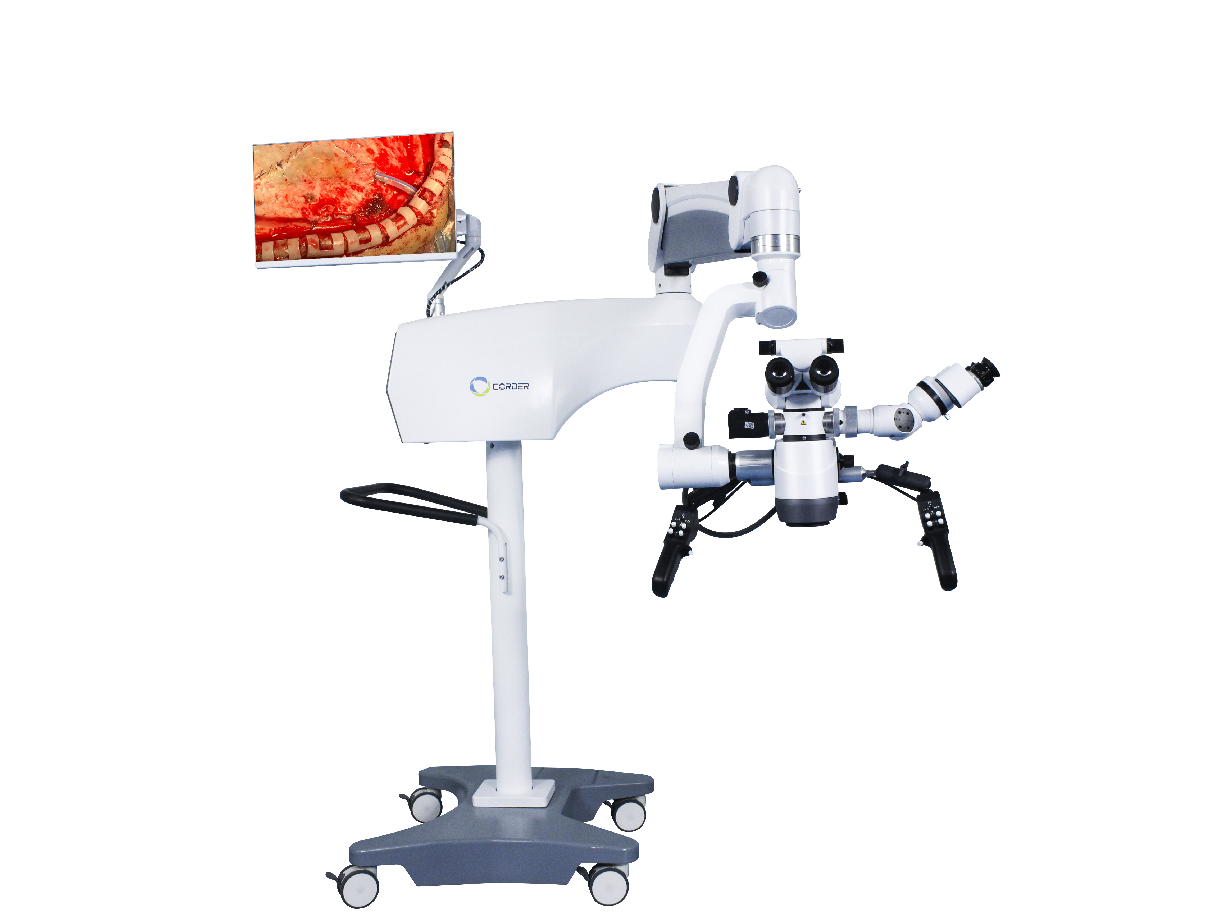
2.脳神経外科における手術用顕微鏡の応用
神経外科の特殊性により、脳神経外科における手術用顕微鏡耳鼻咽喉科や眼科よりもやや遅れて登場し、脳神経外科医はこの新しい技術を積極的に学んでいる。当時、手術用顕微鏡の使用主にヨーロッパで普及した。アメリカの眼科医ペリットが最初に紹介した。手術用顕微鏡1946年にヨーロッパからアメリカに伝わり、アメリカの脳神経外科医が使用する基礎を築いた。手術用顕微鏡.
人間の生命の価値を尊重するという観点から、人体に使用されるあらゆる新しい技術、機器、または器具は、事前に動物実験と操作者に対する技術訓練を受けるべきである。1955年、アメリカの神経外科医マリスは、動物に脳外科手術を行った。双眼手術用顕微鏡南カリフォルニア大学の神経外科医であるクルツェは、顕微鏡下で耳の手術を観察した後、研究室で顕微鏡を用いた手術技術を1年間かけて習得した。1957年8月、彼は5歳の子供に顕微鏡を用いて聴神経腫瘍の手術を成功させた。耳科手術用顕微鏡これは世界初の顕微鏡手術でした。その後まもなく、クルツェは、手術用顕微鏡そして、子供の回復は良好だった。これは世界で2例目の顕微鏡手術だった。その後、クルツェはトラックを使って手術用顕微鏡マイクロサージェリー神経外科手術のためにさまざまな場所へ行き、強く推奨した。手術用顕微鏡他の脳神経外科医へ。その後、クルツェは脳動脈瘤クリッピング手術を実施した。手術用顕微鏡(残念ながら、彼は論文を発表しなかった。)治療した三叉神経痛患者の支援を受けて、彼は1961年に世界初の頭蓋底マイクロ神経外科研究所を設立した。私たちは常にクルツェのマイクロサージェリーへの貢献を記憶にとどめ、新しい技術やアイデアを受け入れる彼の勇気から学ぶべきである。しかし、1990年代初頭まで、中国の一部の神経外科医はそれを受け入れなかった。脳神経外科用顕微鏡手術のため。これは問題ではありませんでした脳神経外科用顕微鏡それ自体は問題ではないが、脳神経外科医たちのイデオロギー的理解に問題がある。
1958年、アメリカの神経外科医ドナギーは、バーモント州バーリントンに世界初のマイクロサージェリー研究・訓練研究所を設立した。初期の頃、彼は上司からの混乱や資金難にも直面した。学問の世界では、脳血栓症患者から血栓を直接摘出するために皮質血管を切開することを常に構想していた。そこで彼は血管外科医のジェイコブソンと動物実験および臨床研究で協力した。当時、肉眼では直径7~8ミリメートル以上の細い血管しか縫合できなかった。より細い血管の端々吻合を実現するために、ジェイコブソンはまず眼鏡型の拡大鏡の使用を試みた。その後すぐに、彼は耳鼻咽喉科手術用顕微鏡研修医時代に手術のために。そこで、ドイツのツァイスの協力を得て、ジェイコブソンは2人操作者用手術用顕微鏡を設計した(学位鏡)血管吻合術では、2人の外科医が同時に手術を行うことができる。広範な動物実験の後、ジェイコブソンは犬の非頸動脈の顕微鏡下吻合術に関する論文(1960年)を発表し、血管吻合の開存率は100%であった。これは、顕微鏡下神経外科および血管外科に関連する画期的な医学論文である。ジェイコブソンはまた、マイクロハサミ、マイクロニードルホルダー、マイクロ器具ハンドルなど、多くの顕微鏡下手術器具を設計した。1960年、ドナギーは、手術用顕微鏡脳血栓症の患者のために。米国出身のロートンは1967年に顕微鏡下で脳の解剖学の研究を始め、マイクロサージェリー解剖学という新しい分野を開拓し、マイクロサージェリーの発展に大きく貢献した。手術用顕微鏡マイクロサージカル器具の改良により、ますます多くの外科医が使用するようになっています。手術用顕微鏡外科手術に関する専門家であり、顕微鏡下手術に関する関連論文を多数発表している。
3.中国における脳神経外科における手術用顕微鏡の応用
日本在住の愛国的な華僑である杜子偉教授は、国内初の神経外科用顕微鏡および関連マイクロサージカル器具1972年に蘇州医科大学附属病院(現蘇州大学附属第一病院脳神経外科)に着任。中国帰国後、頭蓋内動脈瘤や髄膜腫などの顕微鏡手術を最初に行った。神経外科用顕微鏡北京義烏病院脳神経外科の趙亜都教授は、蘇州医科大学の杜子偉教授を訪ね、マイクロサージカル器具の使用法を視察した。手術用顕微鏡上海華山病院の史玉泉教授が杜子偉教授の診療科を視察するために自ら訪問した。その結果、導入、学習、応用の波が起こり、脳神経外科用顕微鏡これは中国の主要な脳神経外科センターで火がつき、中国におけるマイクロ脳神経外科の始まりとなった。
4.顕微鏡手術の効果
使用により神経外科用顕微鏡6~10倍の拡大条件下では、肉眼では不可能な手術も可能になる。例えば、篩骨洞を通して下垂体腫瘍手術を行うことで、正常な下垂体を保護しながら安全に下垂体腫瘍を特定し切除することができる。脳幹腫瘍や脊髄髄内腫瘍など、肉眼では不可能な手術もより良く行えるようになる。王忠成院士は、この装置を使用する前は脳動脈瘤手術の死亡率が10.7%だった。脳神経外科用顕微鏡1978年に顕微鏡を使用した後、死亡率は3.2%に低下した。手術用顕微鏡6.2%であり、1984年以降は、脳神経外科用顕微鏡死亡率は1.6%に低下した。脳神経外科用顕微鏡これにより、頭蓋切開を必要とせずに低侵襲の経鼻経蝶形骨アプローチで下垂体腫瘍を治療することが可能になり、手術死亡率が4.7%から0.9%に低下します。これらの結果を達成することは、従来の大がかりな眼科手術では不可能です。手術用顕微鏡これらは現代脳神経外科の象徴であり、現代脳神経外科において不可欠でかけがえのない手術器具の一つとなっている。
投稿日時:2024年12月9日


