
この記事は歯科手術用顕微鏡の理解を深めるのに役立ちます
歯科手術用顕微鏡口腔医療分野における「スーパールーペ」とも呼ばれる、歯科手術および診断に特化した精密機器です。複雑かつ精巧な構造を駆使し、口腔内の微細な構造を医師に鮮明に提示することで、精密な治療を可能にします。
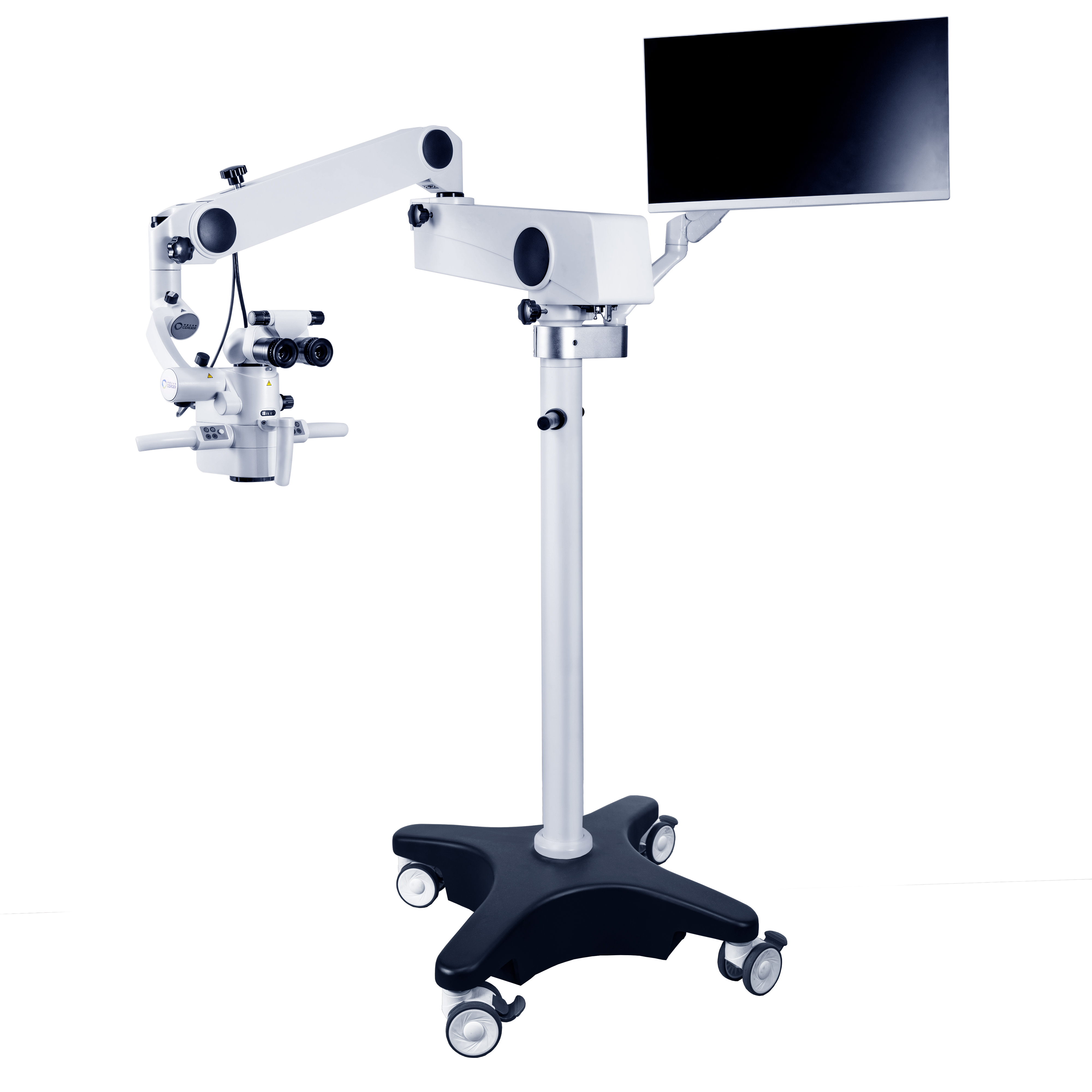
構造的な観点から見ると、歯科手術用顕微鏡主に次の主要コンポーネントで構成されます。
光学拡大システム:これは、顕微鏡カメラのレンズのように、画像の倍率と鮮明度を決定します。現代の歯科手術用顕微鏡通常4〜40倍の間で調整可能で、医師はカメラの焦点距離を調整するのと同じように、手術のニーズに応じて簡単に倍率を切り替えることができます。低倍率(4〜8倍)は、口腔外科手術中に手術領域全体の状態を確認するなど、広い手術視野を観察するのに適しています。中倍率(8〜14倍)は、根管治療、歯周外科手術など、ほとんどの従来の歯科手術のニーズを満たします。高倍率(14〜40倍)では、医師は歯内の根管分岐や象牙細管などの非常に微妙な構造を見ることができ、細かい手術を強力にサポートします。
照明システム:良好な照明は、明瞭な観察の基礎となります。歯科手術用顕微鏡LED冷光源などの先進的な照明技術を採用し、口腔内の手術部位に均一で明るく影のない光を提供します。この照明方法は、従来の光源による熱による口腔組織の損傷を防ぐだけでなく、医師があらゆる角度から手術部位の細部まで観察できるようにし、まるで明るい舞台で演技しているかのように、あらゆる動きをはっきりと見ることができます。
サポートおよび調整システム:このシステムは、手術用顕微鏡、確実に手術用顕微鏡適切な位置に安定して設置され、柔軟に調整可能です。医師と患者のさまざまなニーズに合わせて高さと角度を正確に調整できるため、医師は手術中に最も快適で観察しやすい姿勢を見つけることができます。まるで医師専用の手術台をカスタマイズしているかのようです。
撮影・記録システム:いくつかの高級歯科手術用顕微鏡高解像度カメラと同様の撮像・記録システムも搭載されており、医療用手術顕微鏡リアルタイムで画面に表示されるため、医師は手術中にアシスタントと観察結果を共有しやすく、手術の進行状況の記録や写真撮影も可能です。これらの画像や動画は、その後の症例分析や教育研究に活用できるだけでなく、患者が口腔状態や治療過程をより直感的に理解するのに役立ちます。
の動作原理歯科用顕微鏡光学イメージングの基本原理に基づいています。簡単に言うと、対物レンズと接眼レンズの組み合わせを通して口腔内の小さな物体を拡大します。照明システムから光が照射され、手術部位を照らします。物体からの反射光は、まず対物レンズで拡大され、次に接眼レンズでさらに拡大され、最終的に医師の目または画像装置に鮮明な拡大像を形成します。これは、物体を観察するために虫眼鏡を使用するようなものですが、拡大鏡の拡大効果は口腔外科用顕微鏡より正確で強力なため、医師は肉眼では検出が難しい微妙な詳細を観察できます。
デジタル化、インテリジェンス化、小型化技術の継続的な発展により、歯科用医療顕微鏡機能と性能の飛躍的な向上が期待されます。この技術が大規模病院だけでなく、より多くのプライマリヘルスケア機関や歯科医院にも広く導入され、より多くの患者様に恩恵をもたらすことを期待しています。同時に、手術顕微鏡メーカー研究開発投資を増やし、技術レベルを向上させ、より良いものを製造することができる手術用顕微鏡共同で推進する歯科用顕微鏡業界を新たな高みへと導き、経口薬の発展にさらに貢献します。
投稿日時: 2025年1月20日


